
3D Bioprinting for skin regeneration: Alternative to plastic surgery?
SHRUTI KHANPARA
DR DY PATIL BIOTECHNOLOGY AND BIOINFORMATICS INSTITUTE
Shrutikhanpara16@gmail.com
The field of tissue engineering and regenerative medicine works on creating a functional tissue that mimics the living body tissue. Due to little success in the traditional tissue engineering process, a new approach has been put forth by researchers in which 3D bioprinting is used.
3D bioprinting is an extended application of additive manufacturing that involves a top-down approach of building the tissue in a layer-by-layer fashion thus producing precise geometries due to controlled matter deposition with the help of computer graphics. Stem cells and biocompatible ink is also known as bioinks (biocompatible natural or synthetic material that mimics extracellular matrix to support the proliferation and differentiation of cells) are used for construction that mimics the behavior of living tissue. One of the advantages of bioprinting over the traditional method is that this is a fully automated process, thus giving precise patterning of the cell with the controlled organization.
Bioinks and Cell selection: One of the foremost requirements of bioprinting is bioink. The functionality of the bioprinted tissue is highly dependent on the cell type and bioink composition in terms of rheology, biocompatibility, biodegradation, and antimicrobial activity.
Bioinks are biocompatible materials or hydrogels. Hydrogels are a 3D network of hydrophilic polymer chains having the ability to absorb water. Hydrogels are preferred for 3D scaffolds because of their promising result in the wound healing process by providing a moist environment and a cell carrier. Hydrogels can be natural polymers like collagen, gelatin, alginate, or synthetic polymers like polylactic acid or acrylonitrile butadiene. Hydrogels for bioprinting require to be in liquid form during printing and solid form after printing to maintain desired geometry. To achieve these researchers are trying to use a combination of polymers. For example, alginate/gelatin combination to increase cellular response while co-culturing keratinocyte/fibroblast system in skin tissue.
Appropriate cell selection is another important factor in bioprinting for skin regeneration. Currently, the most common cell types used are fibroblast and keratinocytes which are dermal substitutes and outer epidermis respectively. Since keratinocytes and fibroblast have specific functions and alone could not satisfy the requirement of full-featured skin substitutes it is used in combination to bioprint skin tissue. Nieves Cubo et al. also used plasma along with keratinocytes and fibroblast to mimic human bilayer skin and it was successful in in-vivo clinical trials.
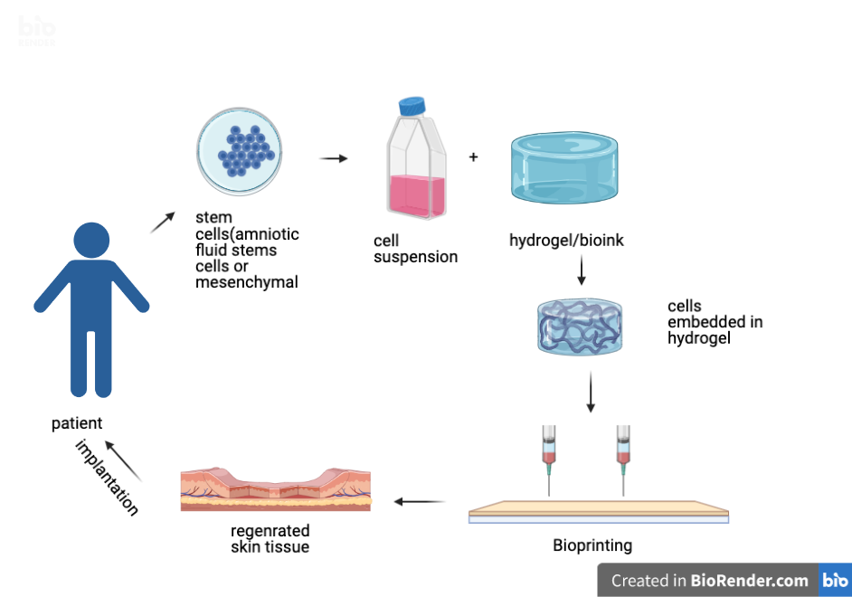
Recently because of the ability of self-renewal and differentiation of stem cells, it is widely used in bioprinting skin tissue. Amniotic fluid stem cells are widely pluripotent and were identified to differentiate in all three cell lineages. These were used in bioprinting for wound healing in mice models and showed successful results with higher wound closer and reepithelialization.
Embryonic stem cells having the capability of proliferating, infinite self-renewal, and generating multiple tissue cell phenotypes show great potential for its use in bioprinting and application in skin regeneration.
Steps involved in 3D skin bioprinting:
It follows three basic steps
Preparatory step: Designing anatomically accurate 3D modeling through computer software by feeding files(patients CT scan imaging or MRI). The preparatory step also includes bioink and cell selection.
Processing step: Actual printing of the tissues by additive manufacturing techniques/3D printers. The bioprinter follows instructions in the code file in order, including instructions to control for the temperature of the extruders, extrusion pressure, bedplate temperature, crosslinking intensity and frequency, and the 3D movement path generated by the program.
Post-processing step: Maturation of fabricated tissue in bioreaction and its structural and functional characterization followed by in-vivo implantation.
Application in 3D skin regeneration:
Laser-assisted bioprinting is used to print keratinocytes and fibroblasts into the 3D model. These cells are embedded in collagen hydrogel to fabricate multicellular skin craft analogous to native archetypes with high cell viability and proliferation. It has been shown to allow precise localization and the ability to mimic tissue-specific functions concerning adhering and gap junctions. It helps to generate a highly organized microvascular network with precise network and branching patterns with high throughput and highly reproducible manner in the mice model. This technology is evaluated in pre-clinical trials and offers much promise in repairing defective tissue in case of brain injuries, tumor resection, skin injury in repairing as well as regeneration.
For wound healing, microextrusion bioprinting is preferred in which amniotic fluid stems cells(AFC’s) and mesenchymal stem cells were suspended in fibrin-collagen hydrogels and were directly bioprinted over the wound site in the mice model. This bioconstruct did show an enhancement in wound closure and reepithelialization. Further proteomic analysis showed that AFC’s secreted growth factors at higher concentration and also induced endothelial migration in-vitro which was an indication that bioprinting AFC could be effective in skin regeneration and curing large wounds and burns.
Limitations:
Although 3D bioprinting is a rapidly developing technique in skin tissue regeneration some challenges still needs to be overcome like:
- Vascularisation and innervation of a printed cell are not achieved. The bioprinted skin might promote wound healing and mimic stratified epidermis but thick vascular dermis is out of reach yet.
- Regeneration of skin appendages like hair follicles is also one of the concerns in 3D bioprinted skin tissue.
- Attention to the printed construct disadvantages like short life span, fast degradation, and severe contraction is also required.
In conclusion, there is a lot to understand and discover to make this technology successful. Once successful, the implementation of this technique can help to obviate the need for the donor in organ transplant and also helps in the restoration of form and function without painful and destructive donor sites.
Reference (Jun-21-A8)
Author Biography: I am a pre-final year student with a major in biotechnology. I have a research interest in biotherapeutics. Aiming to pursue masters in science in the similar field.
